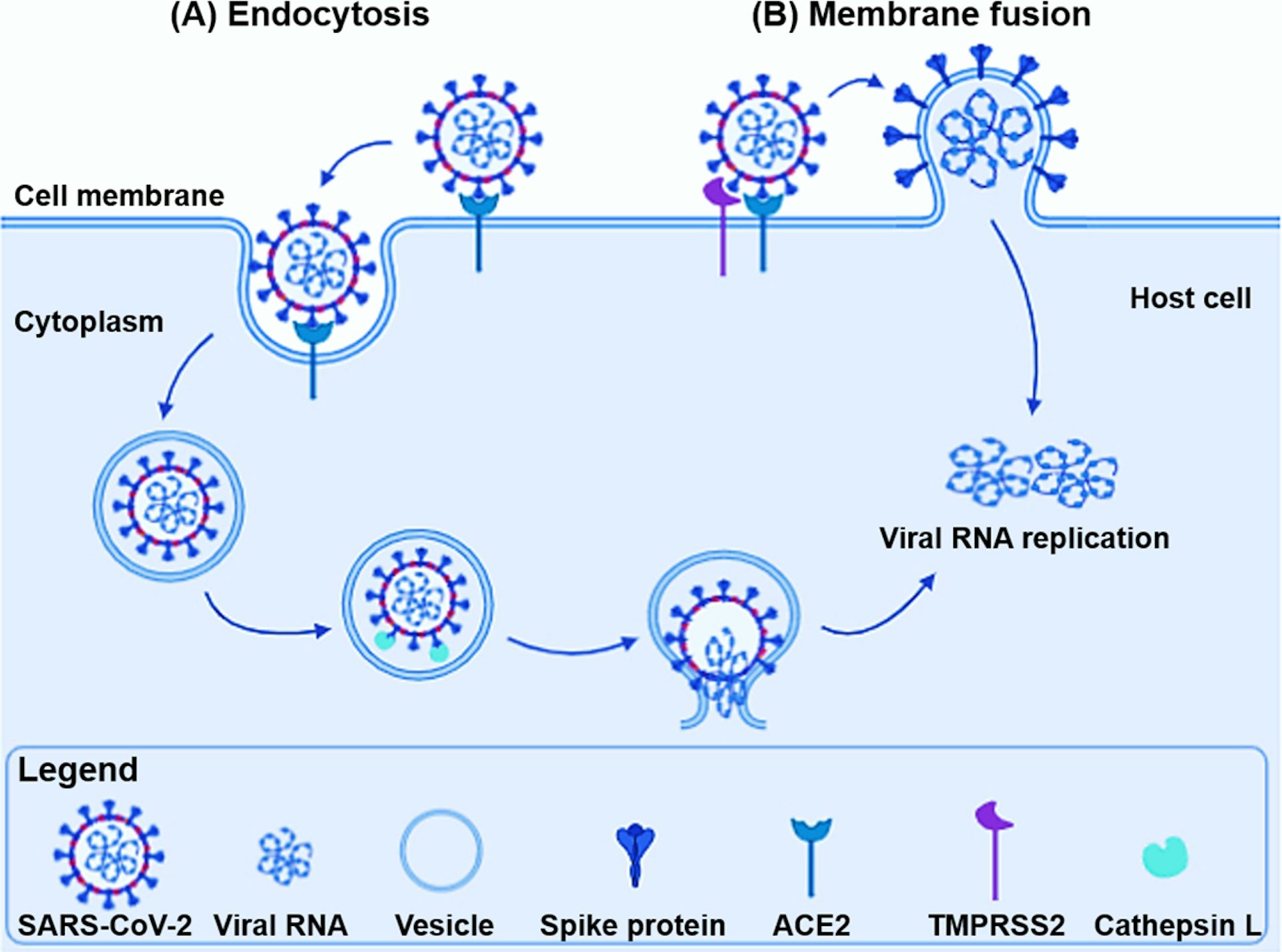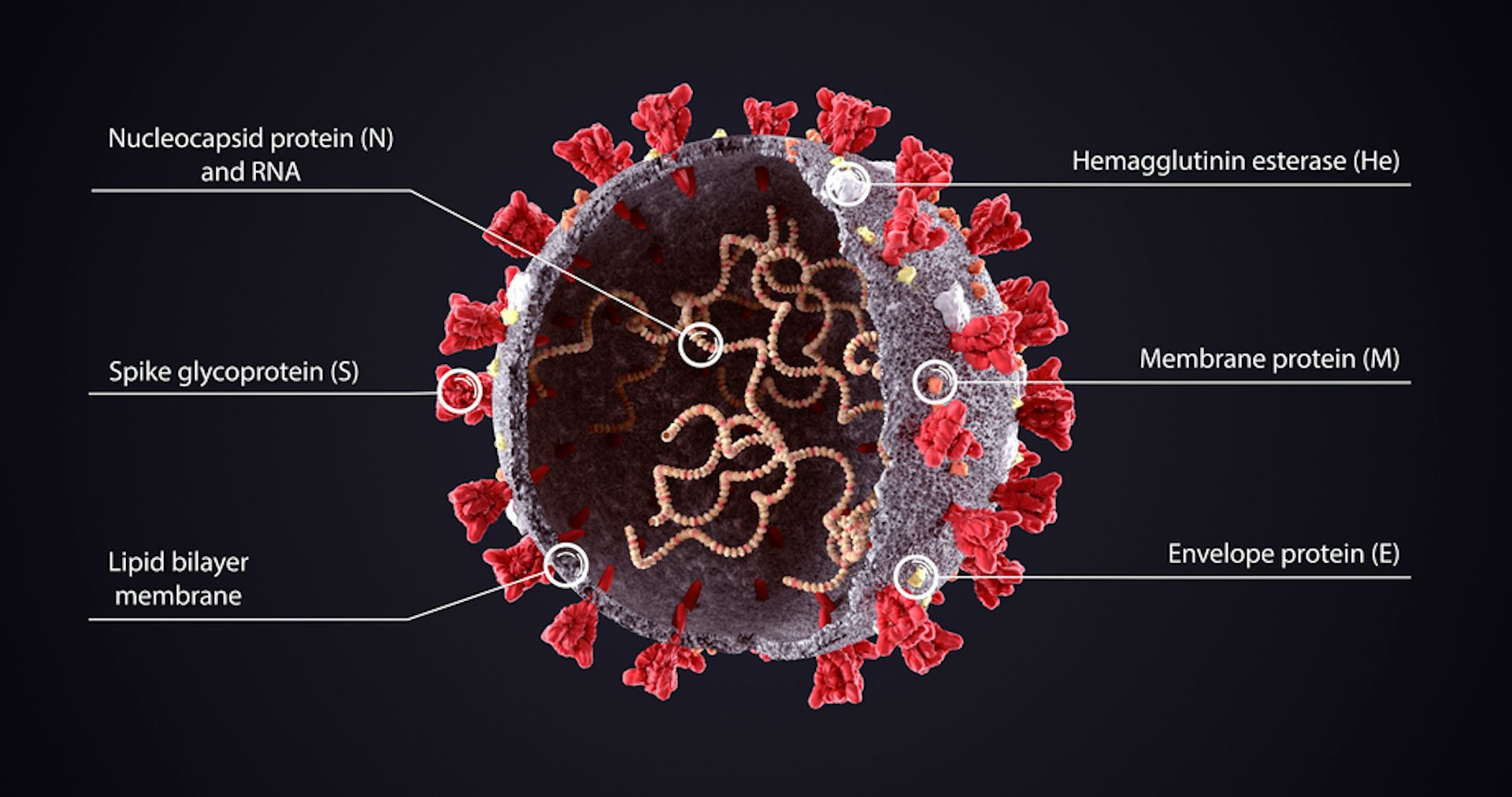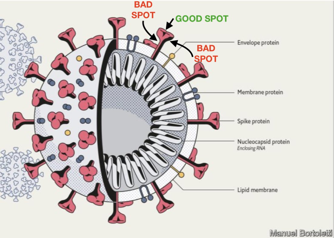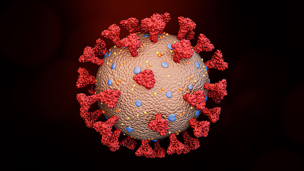

Here is a computer simulation of the movement: As you read this, the molecular jig goes on within your cells to move your eyes, digest your food and help you to think.īy combining molecular dynamics simulations and cryotomography, Sören von Bülow, Mateusz Sikora and Gerhard Hummer at the Max Planck Institute of Biophysics in Germany identified what they likened to the three joints – ‘hip, knee and ankle’ – that give the stalk its flexibility. Nor is the stem rigid, as you would expect of a spike because it is jittering around as a result of thermal energy – the higher the temperature, the more frenetic the molecular movements.Īlmost two centuries ago, the Scottish botanist Robert Brown became fascinated by the incessant zigzag motion of fragments in pollen grains, a seemingly obscure phenomenon that would come to be named after him and provided Albert Einstein with proof that atoms exist.Ĭalled ‘ Brownian motion’ in his honour, this jiggling caused by heat is central to biology and how complex proteins work. A stem anchors the proteins to the virus so, when viewed from above, it looks like a flower with three petals. The name of this protein is misleading since each spike – which consists of three spike proteins – is not ‘sharp’.Įach spike protein links with two others, forming a tulip-like shape. Images courtesy of Peter Coveney, University College London. Vast computer simulations of the protein have been carried out to understand the movement of the spike, such as this one on the Summit supercomputer in Oak Ridge National Laboratory, Tennessee, which included 305 million atoms in the envelope of the virus.ĭifferent views of the spike protein. Here you can see two ways to show the spike protein, either in the form of chains of atoms or by showing the atoms themselves. In the case of titin, which makes muscles elastic, its chemical formula is C 169,719H 270,466N 45,688O 52,238S 911, where C, H, N, O and S refer to atoms of carbon, hydrogen, nitrogen, oxygen and sulphur, respectively. HOW DO WE REPRESENT THE SPIKE?Ĭomplicated biological molecules like proteins consist of vast numbers of atoms. Each spike consists of three copies of the spike protein, S. The spike – there are between 25 and 40 on the surface of each virus – has evolved to stick to proteins on the surface of many cell types that responds to a human hormone that helps maintain blood pressure called angiotensin – hence their name, angiotensin-converting enzyme 2 (ACE2) receptors. By June Almeida and David Tyrrell – Morphology of Three Previously Uncharacterized Human Respiratory Viruses that Grow in Organ Culture, CC0. Thanks to research by David Tyrell, head of the Common Cold Unit near Salisbury, and virus-imaging pioneer June Almeida, the first coronavirus was identified and imaged in the 1960s.

When you use an electron microscope to image the virus – which measures 125 billionths of a metre across – you can see why the coronavirus gets its name: there’s a crown-like haze around the particles which consists of the spikes that it uses to latch on to human cells, the first step required for the virus to invade them. The spikes are around 25 nanometres long– 25 billionths of a metre – and these are big relative to the virus size compared to some other viruses that we are used to looking at.
#VIRAL SPIKE PROTEIN CODE#
The spike is one of just 29 proteins described by the virus’s 30,000 letters of genetic code (by comparison, we have six billion). Proteins, complex chemicals, are the building blocks of living things. WHAT IS THE SPIKE? Image courtesy of MRC Laboratory of Molecular Biology. Here are four representative tomographic slices of SARS-CoV-2 virus particles from infected cells. Cryo-electron microscopy can take images of slices through the virus.
#VIRAL SPIKE PROTEIN SERIES#
The team can also take cryo-EM images of virus samples as they are gradually tilted, resulting in a series of 2D images that can be combined by computer to produce a 3D reconstruction – just like tomography in medicine. Scientists in the LMB study the virus with X rays, or electron microscopes, notably a method called cryo-electron microscopy, cryo-EM, that, like X rays, is now able to ‘see’ individual atoms due to recent improvements. His edited responses are in italic to distinguish them from my commentary. I talked to John Briggs of the MRC Laboratory of Molecular Biology, Cambridge, one of many labs around the world imaging the SARS-CoV-2 virus at the most intimate, atomic level to devise new ways to combat the pandemic.


Roger Highfield, Science Director, describes why it is now the most important protein on the planet. Many COVID-19 drugs, vaccines and tests depend on ‘the spike’.


 0 kommentar(er)
